1.0 Introduction.
Humans and animals have depended on plant as means of food and medicines for decades [1], and there is increase acceptance of medicinal plant in the developed countries [2]. Several cultures still rely on preserved information relating to traditional healing systems, and in Africa and countries like China and India, this has developed into a sophisticated system of diagnosis, medicinal preparations and treatment [3-7]. Medicinal plants represent a considerable source of novel chemicals that could translate to drug development and discovery [3]. In the past, the increased interest in the use of medicinal plants in particular, has been powered by the pharmaceutical industry exploring newer compounds for the treatment of diseases. The World Health Organization (WHO) in recognition of the enormous importance of herbal medicine to primary health care delivery has recommended for actual identification, empirical utilization and development of medicinal herbs for their safety use and efficacy in medical care [3].
Medicinal plants serve as a source of effective therapeutic agents which have been explored as a backbone for the development of some conventional drugs used in the clinics. However, despite the growing scientific evidence of the therapeutic efficacy of medicinal plants [8], studies have indicated that the biological activities of some of these plants are concomitantly associated with adverse effects on the biological system, especially when consumed over a long period of time [9,10]. Therefore, the safety evaluation of medicinal plants intended for use as oral remedies holds an important aspect of drug development from the natural products. The safety of these plants is, however, further jeopardized by the unhygienic and unethical processing, lack of quality controls, appropriate labeling and dosage [11].
Halea ciliata (Mitragyna ciliata Aubrev. Et Pellegr also known as Abura leaves are common among the Yoruba groups of Nigeria for wrapping Kola nuts [12]. It is commonly used in traditional medicine for the treatment of inflammation, headache, malaria hypertension, gonorrhoea, rheumatism, and broncho-pulmonary diseases [13]. Several classes of secondary metabolites produced by Halea ciliata are effective in the wound healing process and have anti-microbial properties [14]. Recently, we reported the antimalarial, anti-inflammatory and analgesic effects of the crude methanol extract of Halea ciliata [15]. However, there is limited published data about its safety and systemic evaluation at high doses of the plant using the oral route, thus its safety profile is needed. In this study, sub-chronic evaluation of crude methanol extract of Halea ciliatawas conducted in Wistar rats
2.0 Materials and Methods
2.1 Collection of plant material
The leaf of Halea ciliata was collected from Ore, Odigbo Local Govt Area, Ondo State, Nigeria. The plant was authenticated by a botanist at Federal Polytechnic Ile-Oluji, Ondo State, Nigeria The Halea ciliata was rinsed under clean running water, air-dried, and pulverized. The powdered sample was stored in an airtight container until it is ready for use.
2.2 Experimental animals
Swiss adult albino rats were obtained from the animal house of the Federal Polytechnic Ile-Oluji. Ondo State, Nigeria. The animals were housed under standard laboratory conditions (Temperature 27 ± 2°C; 70% relative humidity; 12 h daylight/night cycle) and had free access to commercial feed pellets and water [16,17]. The animal study was approved and conducted in strict compliance with the principles governing the use of laboratory animals as laid out by the Federal Polytechnic Ile-Oluji. Ondo State Committee on Ethics for Medical and Scientific Research as contained in the Animal Care Guidelines and Protocol Review of National Institutes of Health Guide for the Care and Use of Laboratory Animals (NIH Publication No. 85-23, 1985).
2.3 Plant extraction
The pulverized Halea ciliata was extracted with methanol using a reflux extractor. Five hundred grams (500 g) of pulverized Halea ciliata was extracted with methanol (2.5 litres) using a reflux extractor. The crude extraction yielded 7.35 % of the extract, which was concentrated, air-dried and stored in a refrigerator at 4 ˚C.
2.4 Sub-chronic toxicity
A total of fifteen albino rats were randomly assigned into three (3) groups of 5 rats each. Single daily doses of 200, and 400 mg/kg BW of the crude extract were administered for 3 weeks to groups 1 and 2. The 3rd group served as the control group and received 0.5 ml of normal saline. All treatments were administered orally once daily using an oral cannula for 21 days. The rats were observed daily for any signs of toxicity, and their body weights were recorded throughout the experimental period.
2.4.1 Collection of blood samples
The collection of blood sample for biochemical and hematological analyses were as described previously [18,19]. Briefly, after the 21th extract administrations, the animals were fasted overnight but had free access to water. The animals were then euthanized using diethyl ether and the blood samples were collected by cardiac puncture into heparinized and non-heparinized bottles and process [20], for hematological and biochemical investigations respectively.
2.4.2 Analysis of serum biochemical parameters
All biochemical analyses were conducted using the Randox Diagnostic kit (Randox Laboratories Ltd, Crumlin, UK). Alanine transaminase (ALT) was analyzed on the principle of the catalytic action of ALT on alanine and α – oxoglutarate to form pyruvate and glutamate [21]. Aspartate transaminase (AST) was measured by monitoring the concentration of oxaloacetate hydrazone formed with 2, 4 – dinitrophenylhydrazine [22]. Serum total protein concentration was estimated based on the principle that cupric ions in an alkaline medium interact with protein peptide bonds resulting in the formation of a colored complex which absorbed maximally at 546 nm [23]. For creatinine assay, creatinine in alkaline solution reacts with picric acid to form a colored complex [24]. The amount of the complex formed is directly proportional to the creatinine concentration. Urea on the other one was estimated spectrophotometrically based on Berthelot’s reaction; urea hydrolysis to ammonia in the presence of urease [25].
2.5 Analysis of hematological parameters
The hematological parameters including hemoglobin concentrations, levels of packed cell volume (PCV) the red blood cells (RBC) counts, white blood cells (WBC) counts, hemoglobin concentration (HB), platelet were estimated using automated hematologic analyzer (Sysmex, KX-21,Japan) as described by Dacie and Lewis [26].
2.6 Statistical analysis
All experiments were conducted with at least three replicates. Analyses were conducted using statistical package for social science (SPSS) version 16 and presented as means ± standard error of the mean. One-way analysis of variance (ANOVA) at p< 0.05 were used for comparing the significant differences between treatment groups.
3.0 Results
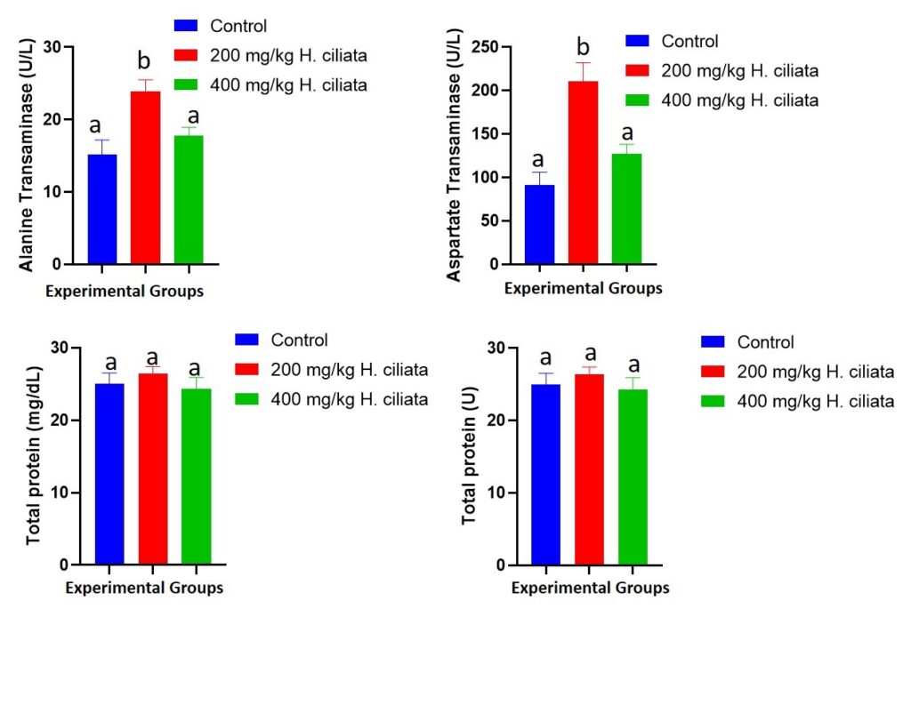
3.1 Effect of sub-chronic administration of crude methanol extract of Halea ciliata on serum activities of some Liver enzymes in wistar rats
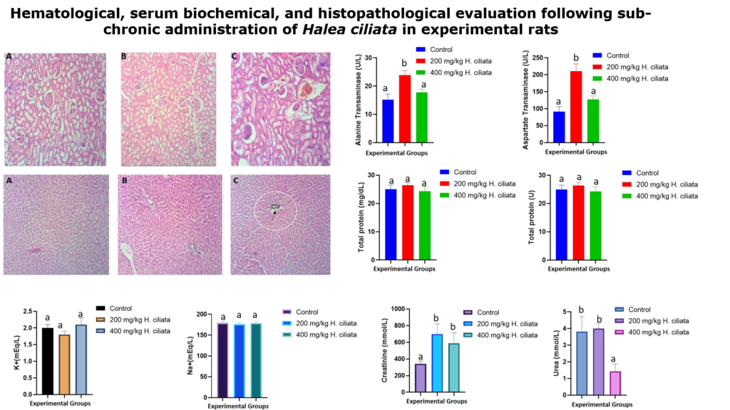
The effect of sub-chronic administration of crude methanol extract of Halea ciliata on serum activities of some Liver enzymes in wistar rats is shown in Figure 1. The activities of ALP and concentrations of total proteins were not significantly (p>0.05) difference among all the groups, including the control. On the other hand, a significant (p<0.05) difference between the 200 mg/kg dose treatment group was observed when compared with the control group in respect to AST, and ALT activities. However, there was no difference between the control group and the highest treatment group of 400 mg/kg, with respect ALT and AST
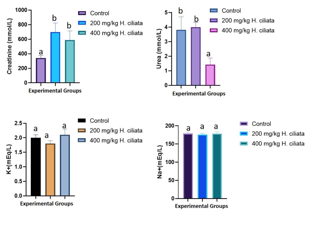
3.2 Effect of sub-chronic administration of crude methanol extract of Halea ciliata on serum biomarkers of kidney function in wistar rats
The effect of sub-chronic administration of crude methanol extract of Halea ciliata on serum urea, creatinine and electrolytes in wistar rats is shown in Figure 2. The concentrations of sodium and potassium were not significantly (p>0.05) difference among all the groups, including the control. On the other hand, a significant (p<0.05) increase in the serum concentrations of urea and creatinine in rats treated with 200 mg/kg bw and those receiving 400 mg/kg bw of the extract was observed when compared with the control group
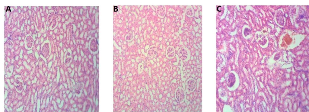
3.3 Effect of the crude methanol extract of Halea ciliata on kidney histology
The kidney section of the control rats shows normal architectural structure of the kidney with well-formed Glomerulus (G) and tubule in the cortical portion. Rats treated with 200 mg/kg bw of the extract demonstrated interstitial edema (IE) coupled with dilation of the lymphatic vessels (DLV) and distortion of the Glomeruli (DG). Similarly, rats treated with 400 mg/kg bw of the extract show a distorted glomeruli (DG), blood congestion (BC), and dilation of the lymphatic vessels (Figure 3).
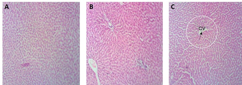
3.4 Effect of the crude methanol extract of Halea ciliata on liver histology
The liver histology in the control rats shows normal liver architectural structure with intact sinusoid channel (s) and hepatocytes (h). However, rats treated with 200 mg/kg bw of the extract shows slight degeneration from the normal architectural structure with necrosis of the sinusoid channels (NC) and slight dilation of central vein (CV) coupled with heamorage while rats treated with 400 mg/kg bw show slight degeneration from the normal architectural structure, central vein with radiating cord of hepatocytes (CV) (Figure 4).
3.5 Effect of sub-chronic administration of the of crude methanol extract of Halea ciliata on haematological parameters of rats
The effects of the of crude methanol extract of Halea ciliataon the haematological parameters of rats is shown in Table 1. The white blood cell (WBC) increased (p<0.05) with increase in dose. Other parameters like the RBC count, PCV, PLT, and HB were not significantly different (p>0.05) across all the groups, including the control.
Table 1: Effect of crude methanol extract of Halea ciliataon hematological parameters in wistar rats
| DOSE (mg/kg bw) | Control | 200 mg/kg bw | 400 mg/kg bw |
| PCV (%) | 34.5±7.07a | 42.8±3.89a | 31.8±7.35a |
| RBC (× 109/L) | 3.47±0.06a | 2.85±0.67a | 4.29±1.97a |
| PLT (× 109/L) | 261.5±30.41a | 217.0±49.50a | 255.0±24.04a |
| WBC (× 109/L) | 5375.0±318.20a | 4950.0±2474.88a | 6050.0±1586.0b |
| HB (g/dl) | 11.51±2.36a | 14.30±1.41a | 10.64 ± 2.50a |
Values are mean ± SEM of 3 determinations. Values along the same column with different superscripts are significantly different (p< 0.05). PCV = Packed Cell Volume, RBC = Red blood cells, WBC = White blood cell, PLT = Platelets.
4.0 Discussion
Halea ciliata have been used traditionally to treat wide varieties of conditions, however, there is limited information and literature reports concerning the toxicity and safety of this plant. The present study therefore evaluated the sub-chronic toxicity of the crude methanolic extract of Halea ciliata in Wistar rats. Adverse effects arising from sub-acute plant administrations manifest on vital organs especially the liver and kidney due to their involvement in the metabolism of the substance [27]. Therefore, evaluation of biochemical parameters holds a pivotal position in assessing the integrity of the liver and kidney following possible assault from repeated administration of plant extract.
The most relevant and widely used biochemical parameters in assessing liver integrity include AST, ALT, ALP, and total proteins [28,29]. These enzymes and proteins were primarily found in the liver and are released in a substantial amount to the serum when the liver’s integrity has been compromised [30,31]. Consequently, 21 days’ administrations of crude methanol extract of Halea ciliata at 200 mg/kg BW significantly upregulated the activities of AST, and ALT when compared with the non-treated control. As mentioned earlier, these upregulations of biochemical indices of liver integrity are secondary events that follow liver assault. The bioactive components in the Halea ciliata crude extract must have interacted and assaulted the liver cells through a yet unidentified mechanism. However, no significant alterations were observed in the activities of ALP and concentrations of total proteins when compared with the normal control group. Similarly, the serum concentrations of ALT, AST, ALP and total proteins in rats treated with 400 mg/kg bw were not significantly different when compared with the normal control, suggesting the safety of the extract when administered orally for a period of 3 weeks at this concentration of 400 mg.kg bw. Thus, the plant could be safely use for therapeutic purpose at this dose.
Urea and creatinine are important excretory metabolites and are widely accepted clinical markers of kidney integrity [32]. Unfortunately, treatments of albino rats with crude methanol extract of Halea ciliata at 200 mg/kg and 400 mg/kg significantly (p<0.05) elevate the serum urea and creatinine concentrations when compared with the control counterpart. This alteration is an indication of a compromised kidney function; the altered protein metabolism could be implicated in the observed trends of urea concentrations following the sub-acute extract administrations. Coherently with our findings, previous studies [19,33-35] have also reported significant alterations in the levels of urea and creatinine following sub-acute dosing of plant extract at a dose of 600 mg/kg BW or above.
The hematopoietic system is a carrier of genetic materials, nutrients, metabolites across the body and thus is consider as one of the most important systems in human and are most susceptible to assaults by toxic substances, including toxic metabolite from plant extracts [36,37]. Fortunately, there was no significant difference in RBC count, PCV, PLT, and HB counts in rats treated with crude methanol extract of Halea ciliata when compared with the non-treated control, suggesting that crude methanol extract of Halea ciliata even at high dose does not mediate erythropoietic tendency of the animals. This is in line with the study of Raji et al. [35] who worked on the acute and Sub-acute toxicity profile of crude extract and fractions of Gymnema sylvestre. Similarly, in line with our observation, Muhamed et al.[38] reported that the hematological profile of rats dosed N. campestris was not different from the control counterparts. However, the significant increase in white blood cell (WBC) counts following treatments with 400 mg/kg BW suggested immune cells by the animals in order to cope with the stress induced by the extract [37].
5.0 Conclusion
In conclusion, administration of crude methanol extract of Halea ciliata at doses of 200 and 400 mg/kg BW significantly altered the serum urea and creatinine in rats. However, rats dosed with the same extracts at 400 mg/kg were devoid of biochemical anomalies in liver serum biomarkers when compared with the control counterpart. This study therefore, suggested the use of Halea ciliata is relatively safe for liver but its effect on kidney markers call for further investigation
Acknowledgement: Not applicable
Authors’ contributions: All authors contributed equally to the manuscript
Competing interests: No conflict of interest
Funding: The authors funded the research.
References
4. Ebbo, A.A.; Sani, D.; Suleiman, M.M.; Ahmad, A.; Hassan, A.Z. Acute and sub-chronic toxicity evaluation of the crude methanolic extract of Diospyros mespiliformis hochst ex a. Dc (ebenaceae) and its fractions. Toxicology Reports 2020, 7, 1138-1144, doi:https://doi.org/10.1016/j.toxrep.2020.08.028.
16. Ndako, M.; Jigam, A.A.; Kabiru, A.Y.; Umar, S.I.; Lawal, B. Polar extracts from Gymnosporia senegalensis (syn. Maytenus senegalensis) root bark, its effects on nociception, edema, and malarial infection. Phytomedicine Plus 2021, 1, 100113, doi:https://doi.org/10.1016/j.phyplu.2021.100113.
19. Lawal, B.; Shittu, O.K.; Oibiokpa, F.I.; Mohammed, H.; Umar, S.I.; Haruna, G.M. Antimicrobial evaluation, acute and sub-acute toxicity studies of Allium sativum. Journal of Acute Disease 2016, 5, 296-301, doi:https://doi.org/10.1016/j.joad.2016.05.002.
20. Shittu, O.K.; Lawal, B.; Alozieuwa, B.U.; Haruna, G.M.; Abubakar, A.N.; Berinyuy, E.B. Alteration in biochemical indices following chronic administration of methanolic extract of Nigeria bee propolis in Wistar rats. Asian Pacific Journal of Tropical Disease 2015, 5, 654-657, doi:https://doi.org/10.1016/S2222-1808(15)60907-0.
33. Olaniyan, J.M.; Muhammad, H.L.; Makun, H.A.; Busari, M.B.; Abdullah, A.S. Acute and sub-acute toxicity studies of aqueous and methanol extracts of Nelsonia campestris in rats. Journal of Acute Disease 2016, 5, 62-70, doi:https://doi.org/10.1016/j.joad.2015.08.006.